Ceriporia Lacerata Mycelium Culture Medium As A Novel Anti-Aging Microbial Material For Cosmeceutical Application
Mar 27, 2023
Keywords: Cistanche lacerata; anti-aging; anti-inflflammation; anti-oxidation; anti-collagenase; filaggrin upregulation; whitening; wound healing
")
Click Here To Know How Cistanche Work For SkinCare
1. Introduction
Among other organs, human skin is a vanguard interface constantly exposed to harmful external stimuli, such as general metabolic reactions, cosmetics, and ultraviolet (UV) irradiation. These factors lead to undesired biochemical byproducts, including reactive oxygen species (ROS), matrix metalloproteinases (MMPs), and advanced glycation end-products (AGEs), which can trigger skin aging, including wrinkles, pigmentation, and loss of skin tone [1,2]. ROS, dangerous oxygen molecules as a pleiotropic physiological signal transmitter, mainly induce cross-linking of collagen and elastin to induce wrinkles and lower skin recovery, which are considered to be the main driving force of aging caused by UV rays and pollution [3]. MMPs are enzymes activated by UV exposure or inflflammation, which contribute to collagen breakdown while inhibiting the formation of new collagen [1]. The formation of AGEs is the result of the reaction of glucose with proteins, including skin collagen, which can contribute to loss of elasticity, wrinkles, inflflammation, inhibition of skin cell growth, and accelerated aging [2]. Since oxidative stress is one of the metabolic factors and pathways most associated with cellular aging, the consumption of functional foods and functional cosmetics with antioxidant activity is increasing signifificantly. Therefore, antioxidants can be beneficial to the human body by directly or indirectly neutralizing ROS, via regulating metabolic pathways and gene expression as the main cellular activation mechanisms. Natural metabolites derived from microbial sources have been used in a variety of applications as a nutritional source and as a skincare agent [4]. Among them, the metabolites derived from fungi contain biologically active ingredients of considerable commercial value including oligosaccharides, exopolysaccharides (EPS), enzymes, peptides, vitamins, and biosurfactants. These metabolites are widely used as the main raw materials for pharmaceuticals, functional foods, and cosmetic products (5-9]. Packed with thousands of antioxidants and anti-inflammatory properties, they have been widely used to fight aging by improving the skin's natural defenses, restoring skin elasticity, increasing moisture content, promoting collagen synthesis, and exhibiting skin-whitening effects (4,5). Besides, these compounds replacing traditional chemical ingredients, are applied in various cosmetic products used to improve the health and beauty of mankind in a safe and detoxifying manner. In particular, the unique biocompatibility, non-toxicity, and functionality of fungal EPS have been widely employed in the cosmetic industry (4). For instance, metabolic compounds such as Schizophyllan, a polysaccharide of p-1,3 P-glucan with p-1,6 branching, extracted from Schizophyllum commune, are known to help with skin anti-inflammatory and UV protection effects (101. Galactomyces ferment filtrate (GFF) has been isolated from Galactomycescandidum to study cosmeceutical effects on the reduction of facial skin pores, skin pigmentation and relieve oxidative stress, while the mechanisms of action underlying EPS of GFF along with its compound information are still unidentified (11]. Cistanche lacerate is a type of white-putrefactive filamentous fungus that plays an important role in bioremediation by breaking down cellulose and lignin (12,13]. The bioactive efficacy of C. lacerata mycelium (CLMculture has been studied to control hyperglycemia levels (14), insulin secretion through cytoprotective effects (15), and insulin signaling through activation of AMP-activated protein kinase (AMPK) and glucose transporter type 4 (GLUT4) (16,17. However, the pharmacological effect of CLM on anti-oxidation and anti-aging is not yet known.
")
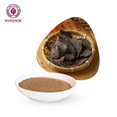
The cultured C. lacerata is composed of microscopic polypores; thus, it looks like white moss (Figure 1a). During the liquid culture process, various secondary metabolites, e.g., exo-metabolites, are generated depending on the environmental conditions (Figure 1b). However, no reports of C. lacerate or CLM on skin care-associated effects exist. Therefore, this study aimed to investigate the antioxidant, wound healing, wrinkle improvement, moisturizing, and whitening effects of CLM on the anti-aging mechanism of skin cells. To the best of our knowledge, this study is the first report to propose a new route for skincare at the cellular level based on the anti-aging mechanism using anti-diabetic ingredients derived from the culture of CLM, an emerging microorganism [14–17].


Figure 1. Green manufacturing method of Cistanche lacerata exo-pharmaceutical substance (CLEPS),including solid culture
(a) and downstream processes (b) such as pre-culture (i), main culture (i), and filtration (i).
2.2. Measurement of Antioxidant Activity2.2.1. 2,2-Diphenyl-1-picryl-hydrazyl-hydrate (DPPH)
Scavenging Activity AssayFree radical scavenging ability of the CLEPS was tested by DPPH radical scavenging photometric assay according to methodology described by Choi et al. (18] andKedare et al. (19. The CLEPS was mixed with 90 mM methanolic DPPH to form final solution concentrations of 0.5, 1, and 5 mg/mL in 96-well plates, which were incubated for30 min at 25 °C and the absorbance (OD) was read in a microplate reader (Multiskan GOThermofisher Scientific, Waltham, MA, USA) at a wavelength of 517 nm. Three independent experiments were performed. Ascorbic acid (AA, 1 mg/mL) was used as the positive control. DPPH inhibitory percent was calculated by following the formula below: DPPH scavenging % = control A0 - sample A1/control A0] x 100, where A1 indicates the absorbance of the sample, while A0 indicates the absorbance of the control (methanolic solution of DPPH).
")
2.2.2. 2,2-Azinobis-(3-ethylbenzothiazoline-sulfonic acid) (ABTS)
Radical Scavenging ActivityDetermination of the free radical scavenging activity of CLEPS solutions was achieved by ABTS radical cation decolorization assay according to a methodology described bKedare et al. (19]. ABTS cation radical generation was accomplished by combining 10mg of ABTS and 2 mg of potassium persulfate in water. The solution was placed in the darkat 25 °C for about 12 h before use. ABTS solution (1 mL) was diluted with 60 mL of methanol, then the CLEPS was mixed with 90 uM methanolic ABTS to form final solution concentrations of 0.5, 1, and 5 mg/ml in 96-well plates. The plates were incubated at 25 ◦C for 30 min and the OD was measured at a wavelength of 734 nm using a Multiskan GO instrument. Three independent experiments were performed. Ascorbic acid (AA,1 mg/mL) was used as the positive control. ABTS inhibitory percent was calculated by following the formula below: ABTS scavenging % = [control A0 − sample A1/control A0] × 100 where A1 indicates the absorbance of the sample, while A0 indicates the absorbance of the control (methanolic solution of ABTS).
2.3. Cell Viability Assay
mounted with 10% fetal bovine serum (FBS) (WelGENE, Gyeongsan, Korea) and 1% penicillin/streptomycin (WelGENE, Gyeongsan, Korea) at 37 ◦C in a 5% CO2 incubator (UP50H, Forma Scientific, Marietta, OH, USA). The cells were incubated at 7 × 10⁴cells/well in a 12-well plate. After confirming the cell adhesion, it was transferred to a serum-free media and the solutions were treated at a working concentration in each well for 72 h. The thiazolyl blue tetrazolium bromide (MTT) (Sigma, St. Louis, MO, USA) solution at a concentration of 100 µg/mL was added to each well and incubated at 37 ◦C, 5% CO2 for 2 h. Then, all the culture medium was removed and 500 µL of DMSO (Duchefa Biochemicals, Haarlem, The Netherlands) was added to each well. The absorbance at 570 nm was measured with an ELISA reader (Epoch, Bio-tek INC, Winooski, VT, USA). The cell culture medium was used as a negative control (control (-)).
2.4. Expression Level of Filaggrin
To showcase how CLEPS influences the skin barrier function, the mRNA expression level of filaggrin was measured by an RT-PCR. HaCaT cell was cultured at 2 × 10⁴ cells/well in a 24-well plate using DMEM medium containing 10% FBS, 1% penicillin, and 1% streptomycin, and then cultured at 37 ◦C, 5% CO2 incubator for 24 h. After removal of the supernatant, each sample was added in DMEM medium excluding FBS for 24 h at 37 ◦C, 5% CO2 incubator. After incubation, the supernatant was removed and the RNA was harvested according to the manual using Easy Blue lysis reagent (iNtRON Biotechnology, Seongnam, Korea). RT PreMix (BIONEER, Daejeon, Korea) was used at 42 ◦C for 60 min and 95 ◦C for 5 min to synthesize cDNA. The PCR primers (Macrogen, Daejeon, Korea) were designed as presented in Table 1 and Western blotting was performed to determine the amount of filaggrin expression.

Lum G (Aplegen, Pleasanton, CA, USA). As a standard internal protein, β-actin (Sigma, St. Louis, MO, USA) was used.
2.5. Melanogenesis Inhibition Test of B16 Melanoma Cells
To find safe doses for melanin inhibition assay and observe the inhibition activity of melanin by the CLEPS sample treatment, B16 melanoma cells (ATCC) at 1 × 10⁵ cells/well were cultured in a 6-well culture plate containing DMEM medium with 10% FBS and 1% penicillin-streptomycin added at 37 ◦C and 5% CO2 condition. A melanin synthesis inducer, α-melanocyte stimulating hormone (α-MSH), was prepared by dissolving it in 10% DMSO at a concentration of 50 µM. After incubating for 24 h, the CLEPS solutions were added and immediately treated with α-MSH (50 nM), followed by additional incubation for 72 h. Next, the cell plates were washed twice with PBS and trypsinized, and then the recovered cells were centrifuged at 5000 rpm for 10 min to remove the supernatant, after which a cell pellet was obtained. The intracellular melanin content was determined according to the method previously reported with minor modifications [20]: melanin was harvested by dissolving it in 2 N NaOH containing 10% DMSO at 60 ◦C for 4 h, which was transferred to a 96-well plate. DMSO (0.1% v/v) was the solvent for the control and the test samples with α-MSH. The absorbance was measured at 475 nm with an ELISA reader.
2.6. Anti-Inflflammation Assay of Nitric Oxide (NO)
medium without LPS (normal group) was used as the negative control.
")
2.7. Anti-Inflflammation Assay of Inducible Nitric Oxide Synthase (iNOS), Cyclooxygenase-2 (COX2), and Tumor Necrosis Factor α (TNF α)
2.8. Synthesis of Collagen and Inhibition of Collagenase
The NHDF cells were cultured and pretreated with CLEPS to examine the efficacy of CLEPS on collagen formation and collagenase inhibition by taking a procollagen type I C-peptide EIA kit (Takara, Kusatsu, Japan) and a human pro-MMP-1 Quantikine ELISA kit (R&D system, Minneapolis, MN, USA), respectively. As a method of measuring the activity level of collagenase, an enzyme that decomposes collagen, an antibody against collagenase (MMP-1) was used, while Phorbol 12-myristate 13-acetate (PMA) (Sigma) was used to activate the expression of MMP-1. 10 ng/mL of TGF-β (Sigma) was dissolved in the culture medium and was used as a positive control, while the culture medium was the negative control.
2.9. In Vitro Wound Healing Assay
To examine the effect of CLEPS on wound healing, HaCaT cells (1 × 10⁴cells/well) were plated into 12-well plates for 48 h and grew to ~100% confluence. The monolayer was wounded using the tip of a sterile 200 µL pipette. Cell debris was removed by washing twice with PBS. These cells were then replaced with fresh 1% serum medium with or without CLEPS (100, 500, and 1000 µg/mL), followed by stimulation for 0–48 h. A control group was used to compare the wound healing properties in the presence and absence of CLEPS. Photomicrographs of the wound closure (cell migration) were taken at 0–48 h of the same wounded areas using the inverted microscope (Zeiss, Jena, Germany). The % change of wound area in pixels was quantified manually for each image using ImageJ Software (v1.52a, National Institutes of Health, Bethesda, MD, USA).
2.10. Statistical Analysis
Bio-functional assays of the CLEPS in each platform were performed in triplicate. The results were expressed as the mean ± standard deviation. Statistical analyses were carried out using ANOVA, followed by Tukey HSD posthoc test or Student’s t-test. These tests were performed using SPSS software (v25, International Business Machines, Armonk, NY, USA) and Microsoft Excel (v1905, Microsoft, USA). A p-value of <0.05 was considered statistically signifificant.
Email:wallence.suen@wecistanche.com Whatsapp +86 15292862950